trigeminal nerve (CN V) is the fifth bilateral cranial nerve.
Additionally, it is the major cranial nerve.
The nerve’s anatomical pathway and the motor, sensory, and parasympathetic functions of its terminal branches will be discussed in this article.
Derivatives of the first pharyngeal arch are connected to the trigeminal nerve.
nerve supply
Sensation
The skin, mucous membranes, and facial sinuses are all innervated by the three terminal branches of the CN V.
Their pattern of distribution is comparable to the supply of spinal nerves in the dermatome (except there is little overlap in the supply of the divisions).
Motor
the mandibular branch of CN V is the only one with motor fibers.
It provides sensory input to the masseter, temporalis, lateral, and medial pterygoid masticatory muscles.
Other 1st pharyngeal arch derivatives supplied by the mandibular nerve include the anterior belly of the digastric, the mylohyoid, the tensor veli palatini, and the tensor tympani.
Parasympathetic Supply
Trigeminal nerve branches carry the post-ganglionic neurones of the parasympathetic ganglia.
(However, take note that CN V IS NOT a component of the PNS supply’s cranial outflow.)
Anatomical Course
Three sensory nuclei—the mesencephalic, main sensory, and spinal trigeminal nerve nuclei—and one motor nucleus—the trigeminal nerve motor nucleus—protrude from the midbrain to the medulla as the trigeminal nerve’s origin.
Within the central nervous system, there are clusters of neurone cell bodies called nuclei (pl nuclei).
The sensory nuclei combine to generate a sensory root at the level of the pons.
A motor root is still being formed by the motor nucleus. Similar to the dorsal and ventral roots of the spinal cord are these roots.
The trigeminal ganglion develops from the sensory root in the middle cranial fossa. A ganglion (pl. ganglia) refers to a collection of the neurone cell bodies outside the central nervous system.
In a depression of the temporal bone, the trigeminal ganglion is situated laterally to the cavernous sinus.
The trigeminal cave is the term for this depression.
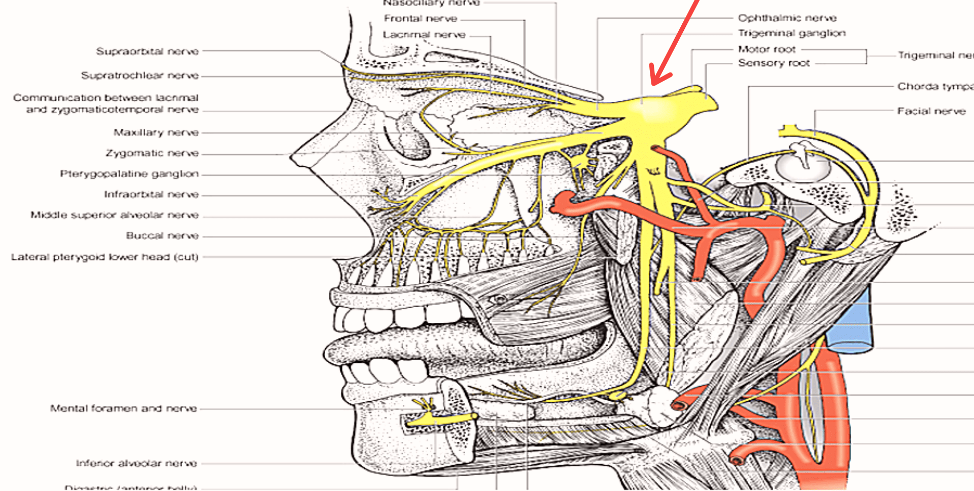
The trigeminal ganglion’s periphery gives rise to the ophthalmic (V1), maxillary (V2), and mandibular divisions (V3).
Along the trigeminal cave’s floor, the motor root connects inferiorly to the sensory root.
It only distributes its fibers to the mandibular division.
The superior orbital fissure and foramen rotundum, respectively, serve as the exit points for the maxillary nerve and ophthalmic nerve from the skull, which move laterally to the cavernous sinus.
The mandibular nerve enters the infra-temporal fossa through the foramen ovale. (Note: Although we are discussing the nerves leaving the cranium, the sensory components can also be said to be entering the cranial cavity, since they are afferent fibres).
we discussing branches of trigeminal nerve in next article